Figure 2.

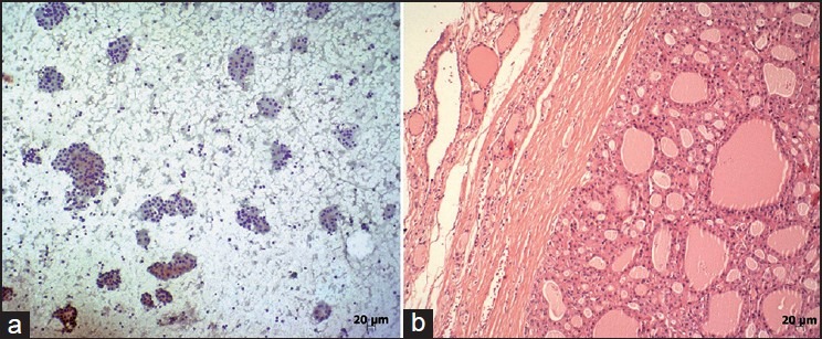
Case with the cytologic diagnosis of “suspicious for follicular neoplasia” (a) (Papanicolaou [PAP], ×100). Histopathologic diagnosis of the same case was Hurthle cell adenoma (b) (H and E, ×100)

Case with the cytologic diagnosis of “suspicious for follicular neoplasia” (a) (Papanicolaou [PAP], ×100). Histopathologic diagnosis of the same case was Hurthle cell adenoma (b) (H and E, ×100)