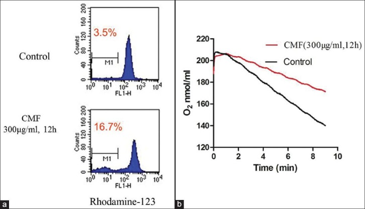
Figure 4.

CMF-induced apoptosis related to mitochondrial dysfunction in K562 cells. (a) Cells treated with CMF (300μg/ml) for 12h were stained with rhodamine-123 to evaluate the mitochondrial transmembrane potential. (b) Alteration of oxygen consumption in K562 cells incubated with CMF (300ug/ml) for 12h compared with control. The images shown here are representative of three separate experiments
