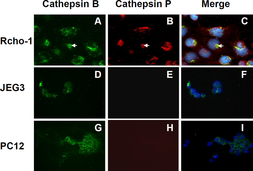
Figure 5. Cellular localization of cathepsins B and P in Rcho-1 cells.
Rcho-1 (A–C), JEG3 (D–F), and PC12 (G–I) cells were fixed, permeabilized, and incubated with rabbit anti-cathepsin P and sheep anti-cathepsin B specific antibodies. Species-specific fluorescent antibodies (Texas red mouse anti-rabbit IgG antibody and FITC donkey anti-sheep IgG antibody) were used to identify the primary IgGs. Cathepsin B is shown as green (A, D, and G) and cathepsin P as red (B, E, and H). Merged images are shown in C, F, and I, with nuclei stained blue with DAPI.

