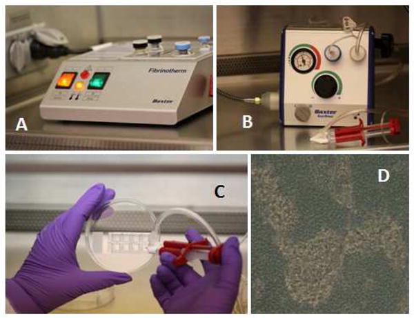
Figure 3.

Composite schematic representation of Adipose Stromal Vascular Progenitor spray deposition. Panel A: Fibrinotherm reagent warmer and mixer. Cells and reagents are warmed to 37° C in this device. Panel B: The Tissomat spray module regulates nitrogen gas used to spray the cells suspended in reconstituted fibrinogen. The Duploject syringe system is in the foreground. Panel C: Cells mixed with fibrinogen sealer protein solution are sprayed onto 8-well chamber slides using the Duploject syringe system. Fibrinogen containing the cell suspension is mixed with thrombin and calcium as they enter the Duploject spray nozzle. Panel D: Light photomicrograph (40 x objective) taken 24 hours after cell/fibrin deposition. Darker areas are the fibrin substrate. Lighter areas represent areas where cells have coalesced within the fibrin, forming refractile clusters.
