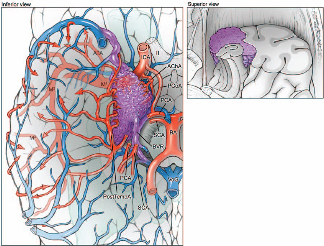
Fig. 3.
Medial temporal AVMs are based on the medial surface (left, inferior view of right hemisphere; right, superior view of coronally transected right temporal lobe). The lesions are supplied by the temporopolar artery, PCoA, AChA, and PTAs from the PCA. These AVMs drain to the BVR and vein of Galen. M1 = sphenoidal segment of MCA; SCA = superior cerebellar artery.

