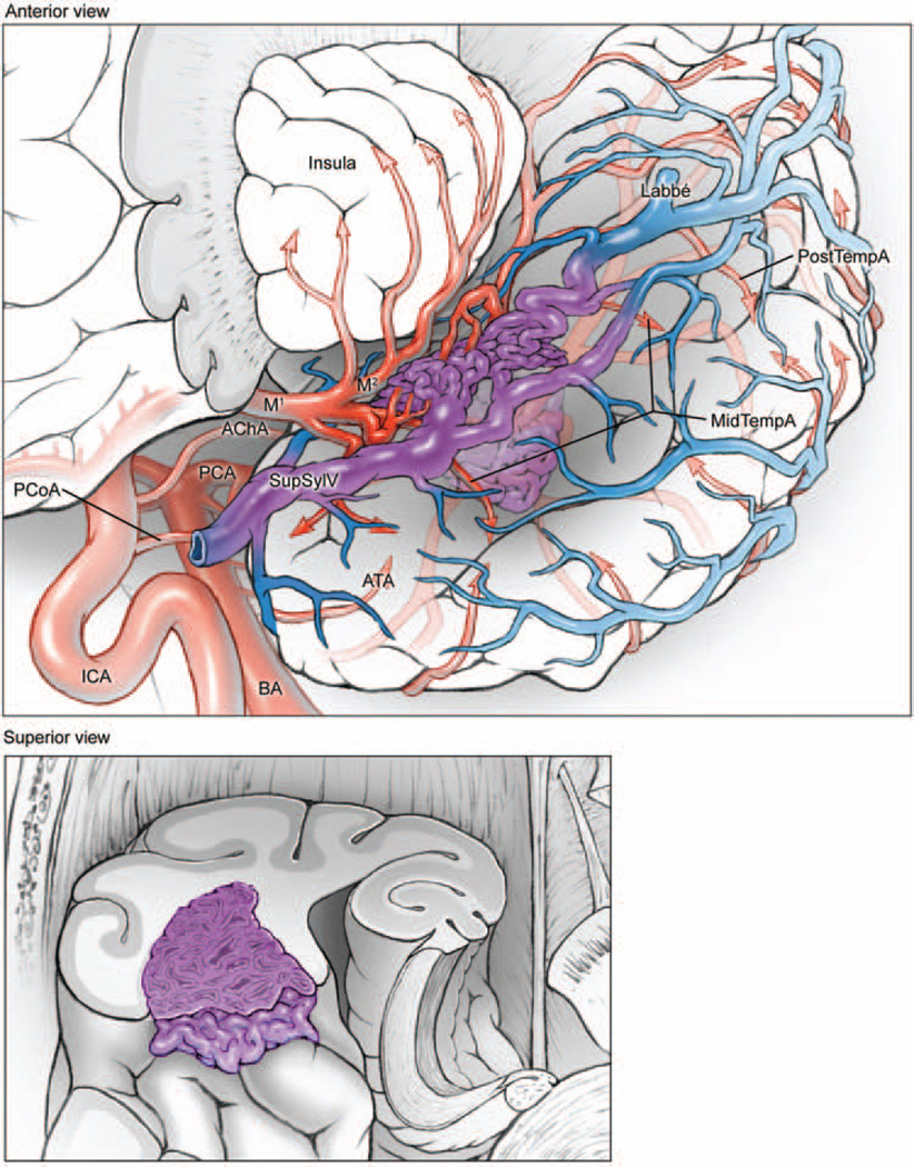
Fig. 4.
Sylvian temporal AVMs are based on the lateral surface of the sylvian fissure (upper, anterolateral view of the left temporal lobe, with lateral left frontal lobe transected parasagittally to expose insular cortex; lower, superior view of coronally transected left temporal lobe). The lesions are supplied by ATA, MTA, and PTA branches from the inferior trunk of the MCA. These AVMs drain to deep and superficial sylvian veins, as well as anterior, middle, and posterior temporal veins on the lateral convexity.

