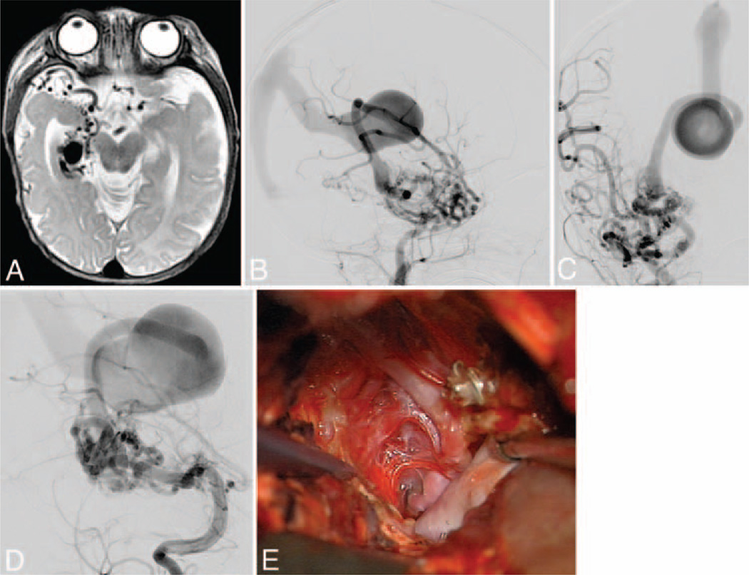
Fig. 8.
Medial temporal AVM. This AVM (Spetzler-Martin Grade III, supplementary Grade I) in a 3-year-old girl occupied the medial temporal lobe (A), as seen on an axial T2-weighted MRI study. It was supplied by AChA and PCoA branches as well as anterior and middle temporal branches of the MCA, as seen on DSAs (right ICA injection; lateral [B] and anteroposterior [C] views). Note the deep venous drainage to the BVR, which has a distal varix and drains into a persistent prosencephalic vein/falcine sinus. Posterior temporal branches from the PCA also fed the nidus (D), as seen on DSAs (right VA injection, anterior oblique view). The AVM was embolized extensively and exposed surgically through a temporal craniotomy, with resection of inferior temporal and occipitotemporal gyri to access the parahippocampus, lateral ventricle, and tentorial incisura. This transcortical dissection exposed the AVM’s lateral margin (E). A large posterior temporal feeding artery filled with coils was transected to access medial feeders from AChA, PCoA, and PCA. The arterialized BVR is seen under the sucker.

