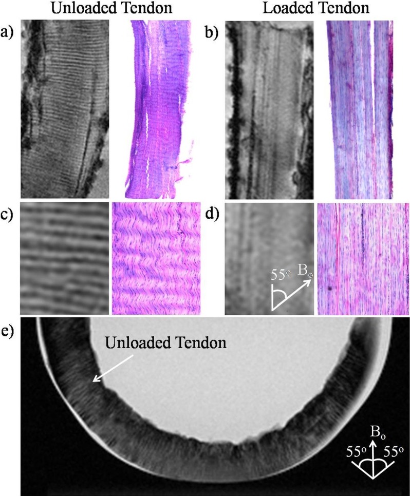
Figure 2.
Illustrations of collagen fiber crimps in magic angle–enhanced MRI and histology, using tendons with and without loading. (a, b) High-resolution T2*-weighted MR images of unloaded ([a]: left) and loaded ([b]: left) tendons oriented at the magic angle at approximately 55° to the main magnetic field (Bo) (repetition time = 3 s; echo time = 5 ms; in-plane resolution = 27 × 27 μm2), and light microscopy of H&E staining in unloaded ([a]: right) and loaded ([b]: right) tendons at ×4 magnification under bright field. As seen in the enlarged views (c, d) of the corresponding images in (a, b), the collagen fiber crimps were revealed as wavy structures by H&E staining in the unloaded tendon, whereas the fibers appeared to be straightened in the loaded tendon. Similar light/dark banding arrangement was observed in T2*-weighted MR images in the unloaded tendon but not loaded tendon. (e) T2-weighted MR image of a U-shaped unloaded tendon positioned at the bottom of a test tube (repetition time = 3 s; echo time = 11 ms; in-plane resolution = 78 × 78 μm2). Magic angle signal enhancement effect could be observed when the tendon tissues were oriented near the magic angle. The collagen fiber crimps were also more clearly distinguished when the tissues were oriented near the magic angle compared with 0° or 90° to Bo.

