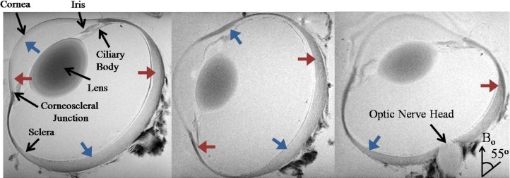
Figure 3.
Representative T2*-weighted MR images of a sheep eye at three different orientations to the main magnetic field (Bo). Note the signal enhancement in the corneoscleral shell when the fiber orientation was near the magic angle at approximately 55° relative to the Bo direction (blue arrows) compared to approximately 0° relative to Bo direction (red arrows) (repetition time = 3 s; echo time = 4 ms; in-plane resolution = 60 × 60 μm2).

