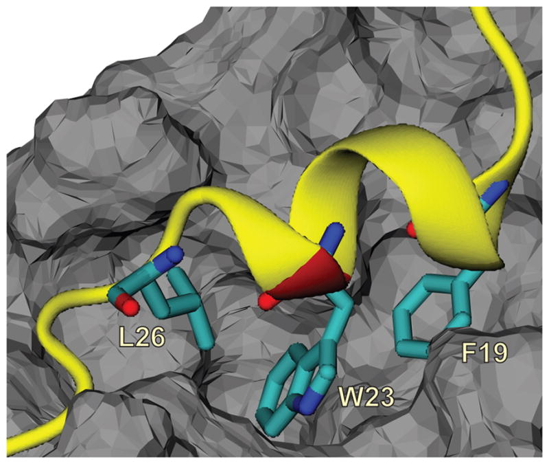
Figure 1.

Structure of the p53TADf-MDM2 complex from PDB ID: 1YCR 7 with three critical residues, F19, W23, and L26 shown in sticks. The p53TADf peptide is presented as a ribbon in yellow with the K24N mutation site colored in red, and MDM2 is shown as grey solid surface.
