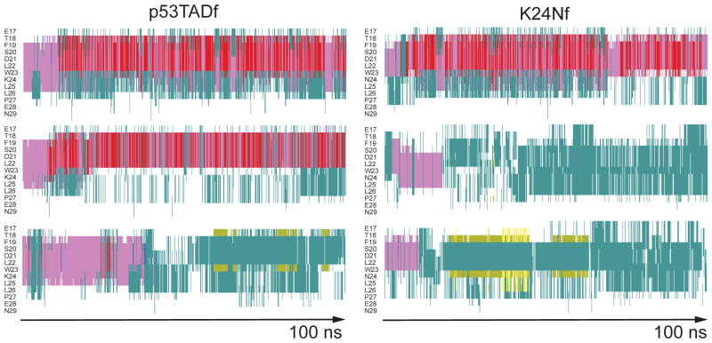
Figure 4.
Secondary structure of unbound p53 fragments from six independent 100 ns molecular dynamics simulations as determined by STRIDE.47 Left column is for p53TADf and right for K24Nf. Color key: alpha helix = purple; 3–10 helix = blue; pi-helix = red; turn = aqua; extended configuration = yellow; isolated bridge = khaki; random coil = white.

