
Figure2. Immunofluorescence staining shows overexpression of Separase in glioblastoma tissue sections.
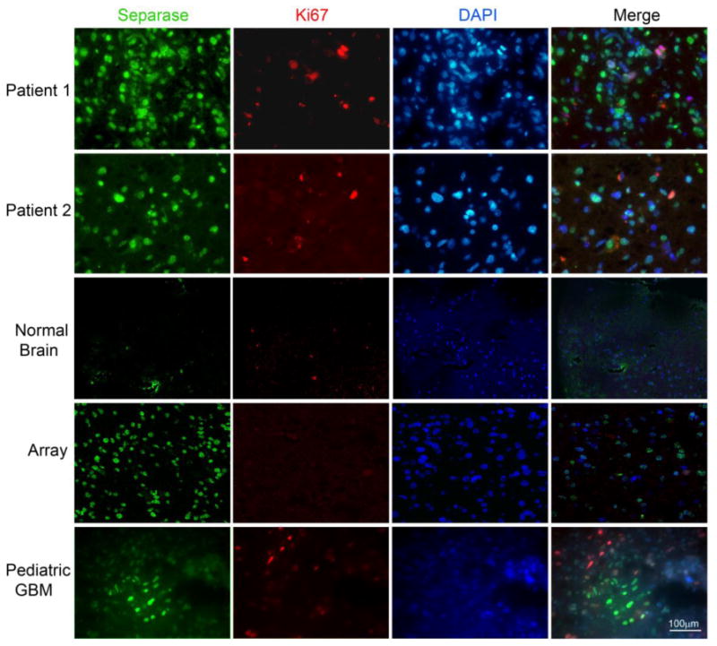
Separase staining (green) showed a higher percentage of positive cells, not all of which were proliferative (shown by Ki-67 staining in red). Normal brain sections stained for the same markers showed only background level of staining. Pediatric glioblastoma shows clusters of positive Separase expression in non-proliferating cells (bottom panel).