
Figure 4. Immunofluorescence images show overexpression of Separase protein in primary glioblastoma cells.
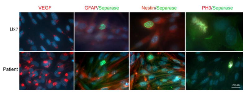
Separase is expressed in nuclei and cytoplasm of cells that are also positive for GFAP and Nestin. Separase expression is detected in nuclei of non-mitotic (PH3 negative) primary glioblastoma cells. VEGF expression is also abundant in these glioblastoma cells.