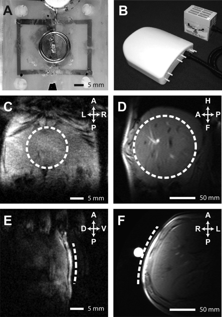
Fig 2.

13C/1H RF coils, and MR images showing coil positioning. (A and B) 13C/1H RF coils employed for preclinical and human studies, respectively. (C-F) Representative hepatic 1H images showing location of 13C surface coils (dashed line) for preclinical (C and E) and human (D and F) studies.
