
Figure 6.
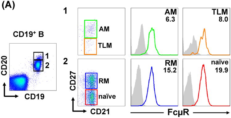
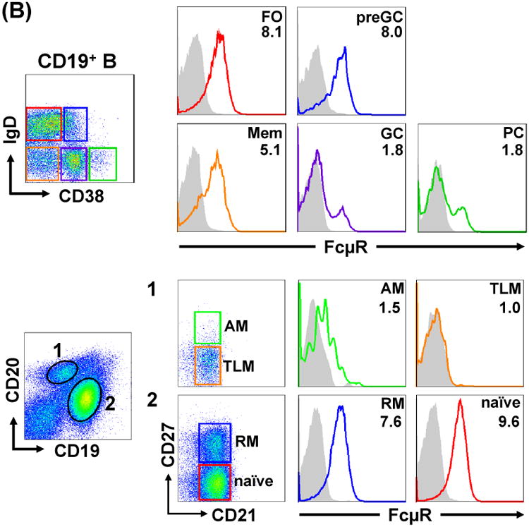
FcμR expression by B cell subsets in blood and tonsils. MNCs from adult blood (A) and tonsils (B) were first incubated with FcγR-blocking reagents and then with biotin-labeled, anti-FcμR (HM14; γ1κ) or isotype-matched control mAb, before developing with PE-SA. PE-stained cells were counterstained with fluorochrome-labeled mixture of four or three mAbs with specificity for: CD19, CD20, CD21 or CD27 (A and B lower panel) or CD19, IgD or CD38 (B upper panel), including fluorochrome-labeled, corresponding isotype-matched control mAbs for background setting. Stained cells were analyzed by BD LSR II (A and B lower) and Accuri C6 (B upper) flow cytometries. Cells in boxes with numbers or different color frames were examined for the reactivity with FcμR-specific (solid lines) or control (shaded histograms) mAb. Numbers indicate the mean fluorescence intensity (MFI) ratios defined as (MFI of anti-FcμR mAb ÷ MFI of control mAb). AM, activated memory; TLM, tissue-like memory; RM, resting memory; FO, follicular; preGC, pregerminal center; Mem, memory; GC, germinal center; PC, plasma cell.