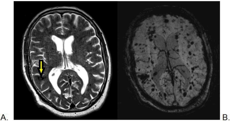
Figure 2.

Magnetic resonance imaging scan of a 78-year-old man with CCM1-CHM. Axial T2-weighted image (A) shows a posterior right temporal lobe typical CCM with reticulated, mixed signal internally and peripheral hemosiderin rim (arrow), and a few small additional small foci of low signal. The axial susceptibility-weighted image (B) is more sensitive for blood breakdown products and shows numerous areas of low signal intensity (dark areas on the image) within small CCMs.
