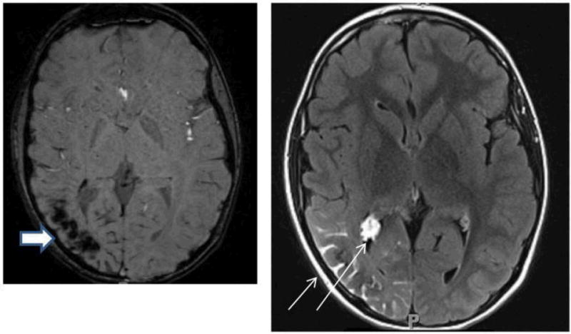
Figure 4.

Axial MRI images from the same individual with Sturge-Weber syndrome showing typical imaging findings of loss of signal on susceptibility-weighted imaging in the right parietal-occipital region, indicating calcification (thick arrow, left panel) and leptomeningeal and choroid plexus enhancement (thin arrow, right panel).
