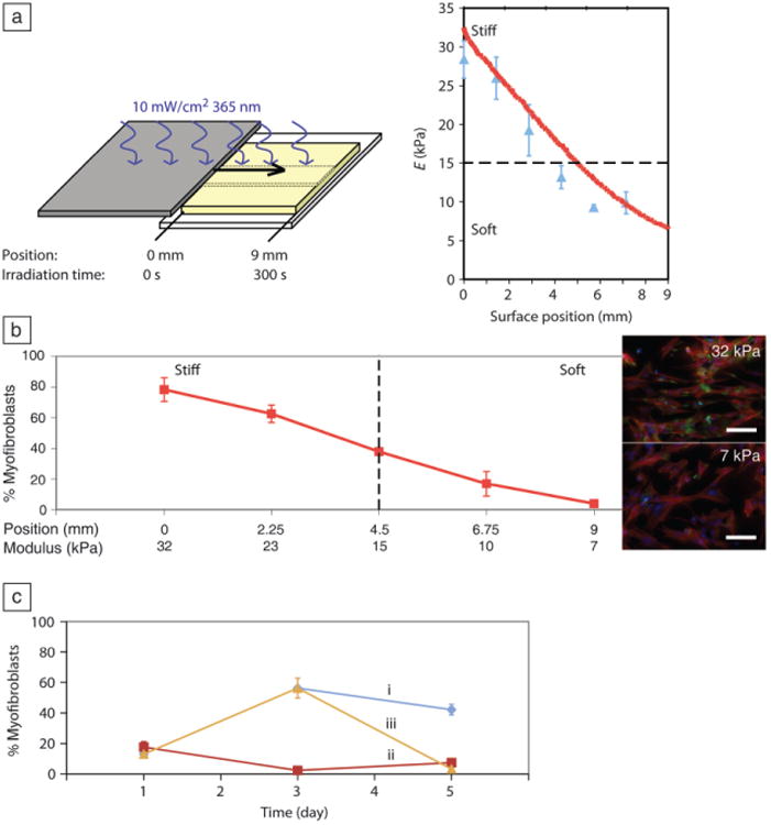
Figure 4.

Valvular interstitial cells (VICs) activation on gradient and stiff-to-soft gels. (a) Modulus gradient across a gel created by moving a mask over the surface during irradiation (left). Modulus (E) measured by atomic force microscopy (blue triangles) and rheometric measurements (red line) as a function of position. (b) The stiff side of the gel promoted VIC activation, whereas cells grown on the soft side remained quiescent on day 3. Percentage of myofibroblasts determined by counting the fraction of cells with α-smooth muscle actin (αSMA) stress fibers, a classic marker for myofibroblasts. Inset: fluorescent images of VICs on stiff (32 kPa) and soft (7 kPa) sides of the gel on day 3, immunostained for αSMA (green), f-actin (red), and nuclei (blue). Scale bar = 100 μm. (c) By softening stiff gels during culture, VICs can be de-activated. (i) 32 kPa “stiff gels,” (ii) 7 kPa “soft gels,” and (iii) 32 kPa-7 kPa “stiff-to-soft gels.” Adapted from Reference 33.
