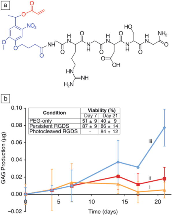
Figure 5.

(a) Structure of the photoreleasable adhesion peptide monomer, arginine-glycine-aspartic acid-serine (RGDS) in black, photolabile moiety in blue, acrylate in red. (b) RGDS presentation maintains human mesenchymal stem cells viability within PEG-based gels (inset table). A chondrogenic marker (glycosaminoglycan, GAG, production) is increased four-fold by day 21 when the adhesive ligand RGD is photoreleased on day 10. (i) PEG-only gels. (ii) Persistently presented RGDS. (iii) Photolytic removal of RGDS on day 10. Adapted from Reference 27. Note: PEG, polyethylene glycol).
