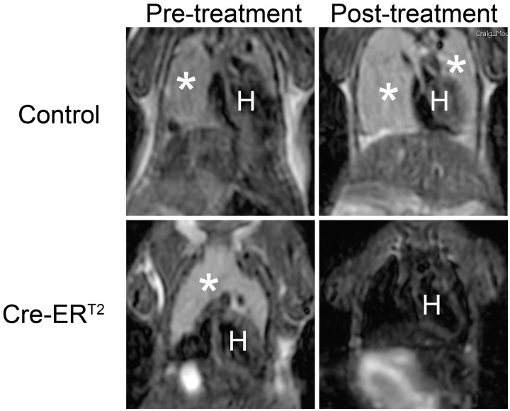
Figure 1. Changes in tumor volume upon tamoxifen treatment in control p53−/− and UBC-Cre-ERT2; p53−/− mice as shown by MRI imaging.
The coronal sections of the thymic lymphoma were shown with tumors labeled with white asterisks. The letter H denotes the location of heart. Post-treatment scans were performed 14 days after starting tamoxifen treatment.

