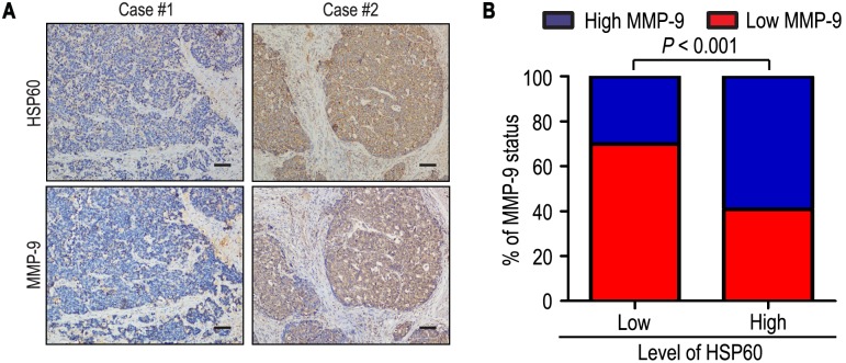
Figure 3. HSP60 and MMP-9 protein levels correlated in 223 gastric cancer tissues.
(A, B) IHC staining for HSP60 and MMP-9 was performed in tumors from 223 gastric cancer patients. Representative examples of HSP60 and MMP-9 staining in serial sections from the same tumor samples are shown in (A), and percentages of samples displaying low or high level of HSP60 relative to MMP-9 level is shown in (B). The scale bar represents 200 µm.

