Figure 1.
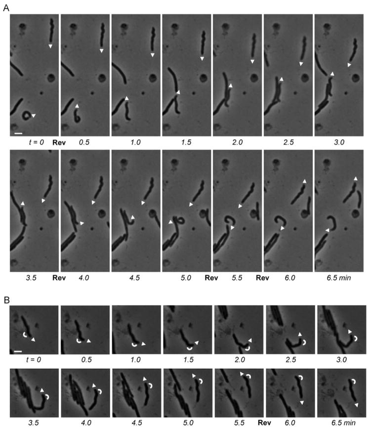
S-motility-deficient cells of Myxococcus xanthus EH302 (ΔpilQ, ΔbacM) gliding over a TPM agar patch 24 h after spotting. Two (A) and one (B) individual crooked cells were marked at the leading pole by arrowheads that indicate the direction of gliding. White bars represent 2 μm; reversals of gliding direction are marked by “Rev”. Arcs in (B) indicate the orientation of the bend near the end of the tracked cell. Note, that the zigzag-shaped cells maintain their orientation during gliding rather than changing into their mirror image (see also corresponding Movies 1A and 1B).

