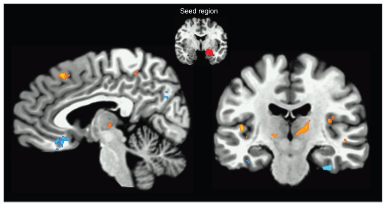
Fig. 1.
Right amygdala seed functional connectivity. The statistical map illustrates areas that showed increased positive coupling with the amygdala (orange) and decreased coupling with the amygdala (blue) during anticipatory anxiety as compared with safety. Areas with significant increases in positive coupling include the medial prefrontal cortex, insula, thalamus and basal ganglia. Areas with significant decreases in coupling include the ventromedial prefrontal cortex, inferior temporal gyrus and precuneus. Sagittal view x = −5; Coronal view y = 18. Images are in Talairach space, neurologic convention, and thresholded at q < 0.05, false discovery rate–corrected.

