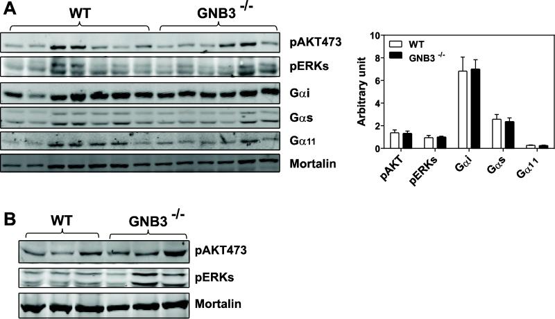
Figure 8. Gβ3 deficiency does not affect GPCR signaling or Gβ subunit expression.
Immunoblot analysis of the levels of phosphorylated AKT473 and ERKs (A, B), or Gαi, Gαs, and Gα11 (A) in the atrium (A) and ventricle (B) of the wild-type (WT) and GNB3−/− mice. Mortalin was used as a loading control. Each lane represents extracts from individual mouse. Quantification of protein expression (normalized by mortalin) in the atrium is also shown (A).

