
Figure 3. Cellular contact through OX40L plays a key role in the ILC2 and CD4+ T cell interaction.
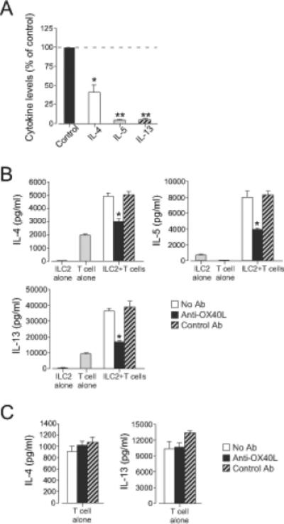
(A) ILC2s (105 cells/well) and CD4+ T cells (2×10⁵ cells/well) were cultured in the Transwell® system for 4 days. Cytokine levels in the supernatants were analyzed by ELISA. Data were normalized to the values from co-culture without the Transwell® system as 100% (control). *, p<0.05; **, p<0.01 as compared to the control. Data (mean±SEM) are a pool of three experiments. (B) ILC2s (104 cells/well) and CD4+ T cells (2×10⁴ cells/well) were stimulated with anti-CD3/CD28 with or without anti-OX40L (10 μg/ml) for 4 days. *, p<0.05 versus no antibody. (C) CD4+ T cells (2×10⁴ cells/well) were stimulated with anti-CD3/CD28 with or without anti-OX40L (10 μg/ml) for 4 days. Data (mean±SEM, n=3) are representative of five experiments.