
Figure 4. ILC2-derived IL-4 plays a key role in the ILC2 and CD4+ T cell interaction.
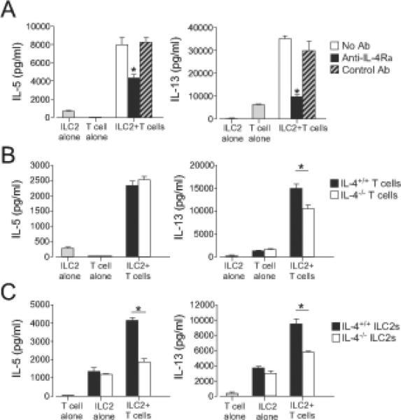
(A) ILC2s (104 cells/well) and CD4+ T cells (2x10⁴ cells/well) were stimulated with anti-CD3/CD28 with or without anti-IL-4Rα(10 μg/ml) for 4 days. Cytokine levels in the supernatants were analyzed by ELISA. *, p<0.05 versus no antibody. (B) ILC2s from WT mice and CD4+ T cells from WT or Il4−/− mice were cultured alone or together for 4 days. *, p<0.05 between the groups indicated by horizontal bars. (C) CD4+ T cells from WT mice and ILC2s from WT or Il4−/− mice were cultured alone or together for 4 days. *, p<0.05 between the groups indicated by horizontal bars. Data (mean±SEM, n=3) are representative of three experiments.