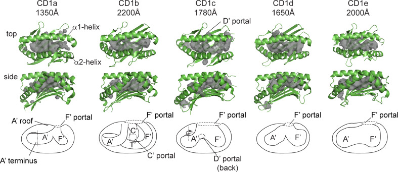
Fig. 2.
CD1 isoforms each have unique-antigen binding groves and capacities. CD1 isoforms have differing groove architecture, as revealed by crystal structures of CD1 with ligands: CD1a with dideoxymycobactin, CD1b with C55 glucose monomycolate, CD1c with mannosyl-phosphomycoketide, CD1d with α-galactosyl-ceramide, and CD1e alone. CD1 structures are rendered in green with cavity surface highlighted in gray and schematic of cavity [figures were generated from RCSB protein data bank files for 1XZO (CD1a), 1UQS (CD1b), 3OV6 (CD1c), 2PO6 (CD1d), and 3S6C (CD1e)]

