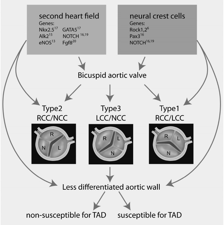
Fig. 2.
Overview of our hypothesis on the developmental origin of the bicuspid aortic valve and aortic wall abnormalities Figure 2 provides a schematic presentation of the aortic bicuspidy valve types based on the valve leaflet orientation and the position of the raphe. Type 1: with fusion between the right coronary cusp (RCC) and left coronary cusp (LCC). Type 2: with fusion between the RCC and non-coronary cusp (NCC). Type 3 with fusion between the LCC and NCC. Furthermore an overview of our hypothesis on the developmental origin of the bicuspid aortic valve and aortic wall abnormalities is provided. Previously identified genetic mutations in mice (Nkx2.5, Alk2, eNOS, GATA5, NOTCH, Fgf8, Rock1,2 and Pax3) and in human (NOTCH) resulting in bicuspid aortic valves (BAV) are indicated in this figure. These genetic defects can be subdivided in either second heart field (SHF) or neural crest cell related. SHF and neural crest cells both contribute to the vascular smooth muscle cells in the ascending aorta as well as to the cells involved in semilunar valve formation. The SHF most probably also contributes to the endocardial cells of the cardiac outflow tract. Therefore these early developmental defects can cause bicuspidy, but also explain the less well differentiated aortic wall seen in all patients with a BAV. However, not every patient with BAV has increased susceptibility for aortopathy. Therefore an additional factor needs to be identified to recognise patients with increased vulnerability for aortic complications

