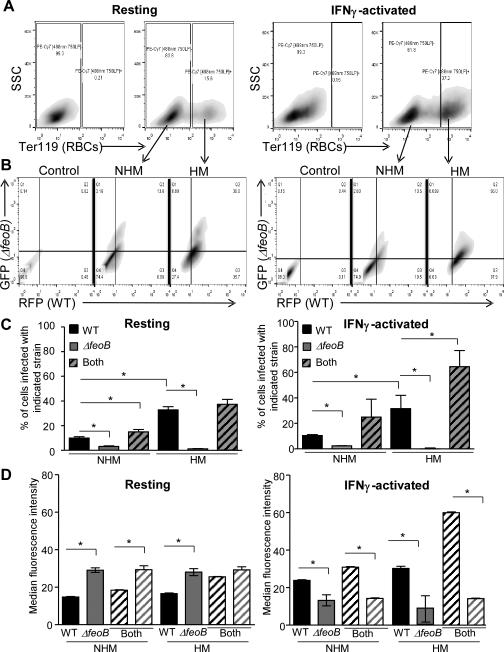
Figure 8. FeoB is required for replication upon mixed-infection in hemophagocytes.
BMDMs were resting (A-D, left panels) or activated with IFNγ (A-D, right panels), incubated with erythrocytes for one hour, and then inoculated with a 1:1 mixture of WT-RFP and ΔfeoB-GFP strains. At 18 hours post-infection, cells were fixed and stained with anti-Ter-119 and analyzed by flow cytometry. A) Gating scheme for non-hemophagocytic (NHM) and hemophagocytic (HM) populations. B) Gating scheme to identify RFP- and GFP-positive BMDMs. C) Percentage of BMDMs that were positive for WT (RFP, black), ΔfeoB (GFP, gray) or both (striped). D) Median fluorescence intensity of WT and ΔfeoB of singly and co-infected BMDMs from (C). Mean and SD of a representative experiment is shown. p-values were determined as described in the methods. p < 0.05 (*) vs. WT, n ≥ 3.

