
Fig. 6.
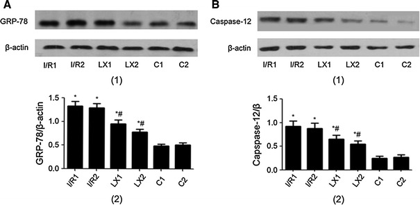
Protein levels of GRP-78 and caspase-12 of cardiac tissues from rats of different groups at T 2 time points. Samples were collected immediately after I/R procedure was completed, and Western blotting was performed as described in Materials and methods. β-Actin was used as a loading control. The lanes shown are representative blots of two independent experiments. A GRP-78 (1, upper) Western blotting; (2, lower) Quantitative data of Western blot. B Caspase-12 (1, upper) Western blotting; (2, lower) Quantitative data of Western blot. * P < 0.05 for comparisons of I/R1 and LX1 groups with C1 group, P < 0.05 for comparisons of I/R2 and LX2 groups with C2 group; # P < 0.05 for comparisons of LX1 group with I/R1 group, P < 0.05 for comparisons of LX2 group with I/R2 group