Fig. 1.
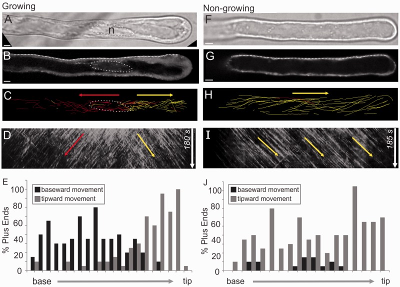
Microtubule growth polarity corresponds to nuclear position in root hairs. Arabidopsis root hairs expressing EB1b–GFP in growing (A–E) and non-growing (F–J) hairs. (A and F) Differential interference contrast (DIC) images showing the nuclear position (circle). The nucleus is to the left of the imaged area in F. (B and G) Midplane confocal slices showing the nuclear position (circle). The nucleus is to the left of the imaged area in G. (C and H) Paths of manually tracked EB1b–GFP dots colored red for baseward or yellow for tipward movement. The nuclear position is shown with a dotted outline. (D and I) Kymographs corresponding to the midplane line drawn on a root hair. Arrows indicate dominant growth directions. (E and J) Histograms showing percentages of baseward vs. tipward MT growth polarities along the length of the root hairs. Scale bars, 5 µm.

