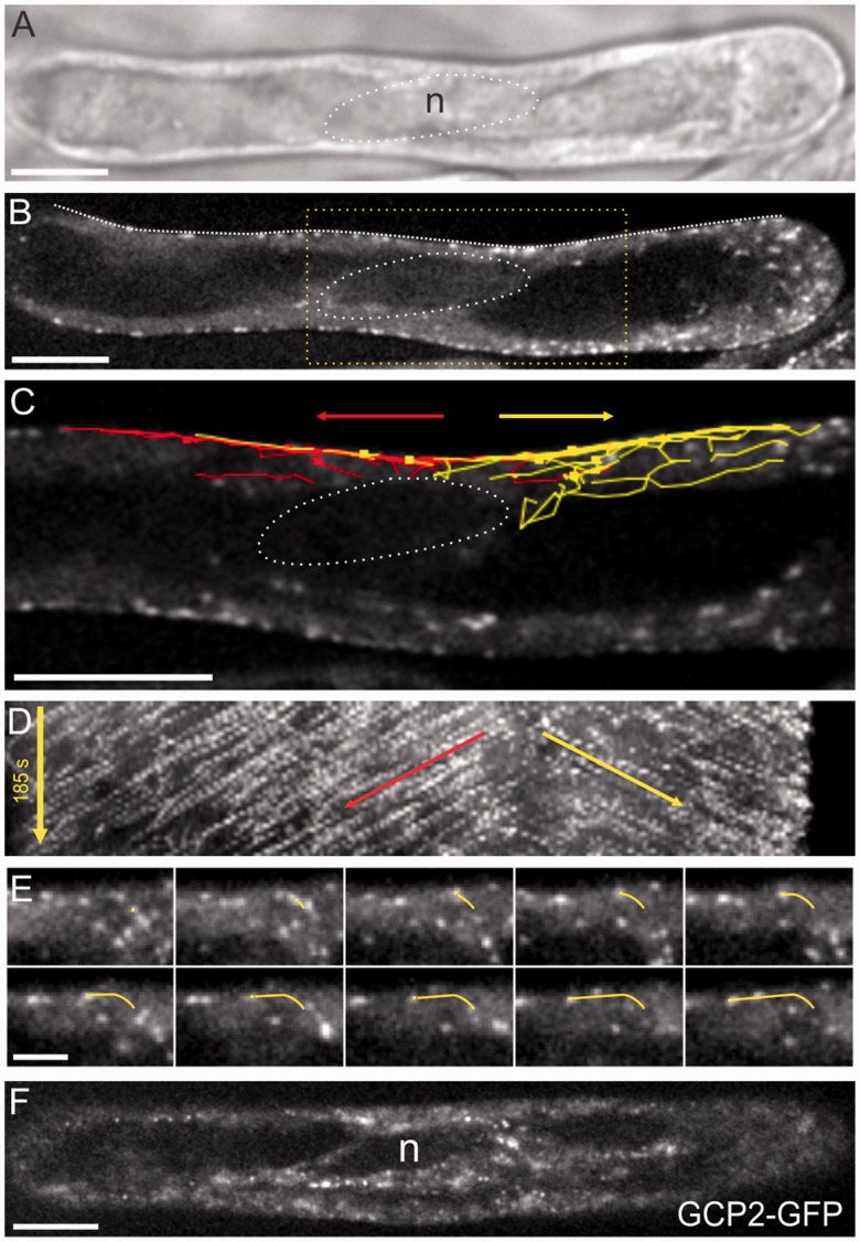
Fig. 2.
Microtubules initiate from the nucleus in root hairs and enter the cortex in two directions (A) DIC image showing the nuclear position (circle). (B) Single EB1b–GFP midplane image showing the nuclear position (dotted circle). (C) Higher magnification of the boxed region in B, showing paths of manually tracked EB1b–GFP dots overlaid on a single time point entering the cortex in two directions above the nucleus. Red is baseward movement and yellow is tipward movement. Arrows indicate dominant growth directions. (D) Kymograph corresponding to the line drawn on the root hair in B. Arrows indicate dominant growth directions. (E) Montage from time series tracking a single EB1b–GFP as it enters the cortex and then grows along the cortex. (The yellow line is shown for reference of the track.) Intervals are 5 s between frames; total time is 50 s. (F) GCP2–3×GFP is localized to the nuclear surface and surrounding the cytoplasmic strand. Shown is an image of the confocal midplane of a root hair. Scale bars, 10 µm for all panels except E, which is 5 µm.

