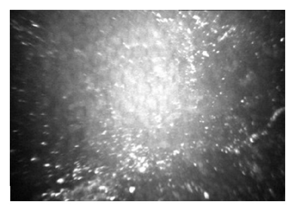
Figure 2.

In vivo confocal microscopic image of a descemetic (D-DALK) interface. One month after surgery, a moderately hyperreflective and homogeneous layer, adjacent to the endothelium, was generally observed. Bright microdots are clearly visible in this case.
