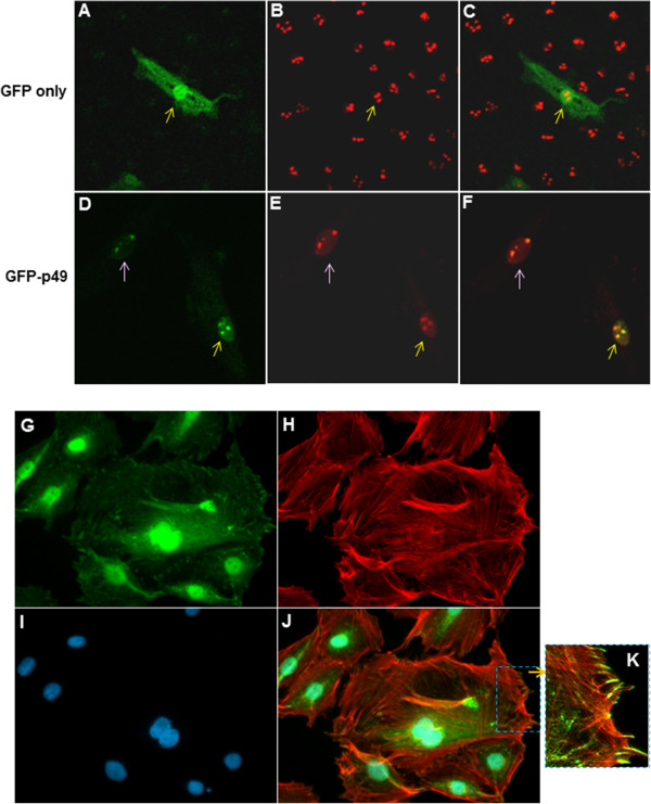
Figure 2.

Intracellular distribution of p49/STRAP proteins. A-F: confocal microscopy revealed that p49 protein co-localized with nucleolin within the nucleolus in H9C2 cells. A. GFP protein is distributed in both cytoplasm and nucleus in GFP control plasmid transfected cells. B. Anti-nucleolin antibody was used to stain the nucleolus. Red fluorescence showed the nucleolus. C. Overlap of images of A and B. D. Transfected GFP-p49 protein was mainly distributed in nucleus and concentrated as dots within the nucleus. E. Anti-nucleolin antibody was used to stain nucleolin which is biomarker of nucleolus. Red fluorescence showed the nucleolin were stained as red dots. F. Overlap of images D and E, revealing GFP-p49 protein (in green) and nucleolin (in red) co-localized as yellow dots in the nucleolus. G-K: The distribution of endogenous p49 protein in H9C2 cells. The figures here revealed the abundance and the distribution of endogenous p49 protein in cells under normal culture condition. G. The endogenous p49 protein is widely distributed in the cell, but concentrated in the nucleus. H. Actin fiber was stained with rhodamine-phalloidin. I. Nuclei were stained with DAPI. J. Overlap of images A, B and C, which indicated that the p49 protein was in close proximity to the actin fiber. K. Digitally enhanced image showing that the p49 protein appeared to form a “tail” that was co-located at each end of the actin fibers, close to their attachment sites to the inner wall of the cell membrane.
