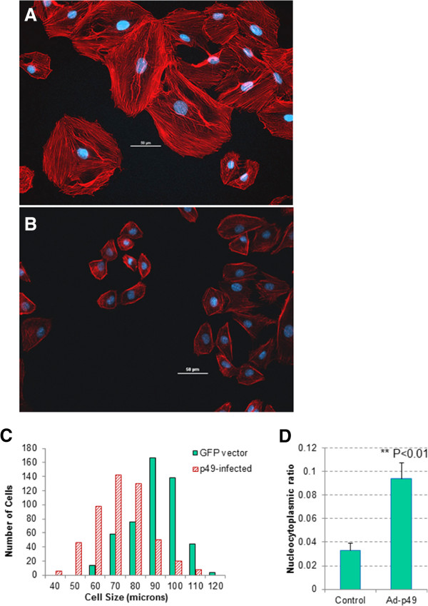
Figure 4.

p49-adenovirus treatment reduced the actin fiber structure and intensity, and changed the cell morphology in term of cytoskeletal structure, cell size, cell shape and nucleocytoplasmic ratio. A. Control H9C2 cells were infected with GFP-control adenovirus and stained with Phalloidin and DAPI for visualization of F-actin and nuclei, respectively. Control cells showed relatively uniform and normal morphology for H9C2 cell line. Nuclei occupied a small, centrally located region of the cytoplasm. Actin fibers were dense and showed uniform network throughout the cytoplasm of the cell. B. P49/STRAP adenovirus infected H9C2 cells were stained with Phalloidin and DAPI for visualization of F-actin and nuclei, respectively. Overexpression of p49/STRAP in H9C2 cells showed smaller overall cell size, along with less uniform cellular shape compared to that of the control. Overall expression of actin was decreased, with significantly less uniformity among visualized fibers. Nuclei of p49/STRAP overexpressed cells were observed to occupy a much greater percentage of the cytoplasm and peripheral location in the cell. C. Histogram of cell size and numbers in H9C2 cell samples that were infected with p49-adenovirus versus control adenovirus. The cell size and number were measured under microscope and multiple fields were examined. The overall cell size in p49 adenovirus-infected samples was smaller versus control adenovirus-infected cells (n = 500, p <0.05). D. Histogram showed the nuclear to cytoplasmic ratio among control H9C2 cells infected with GFP only and adenovirus p49/STRAP infected cells. Data indicated that overexpression of p49/STRAP resulted in a significant increase in the nuclear to cytoplasmic ratio compared to that of the control (p < 0.05, n = 50). These results suggest that treatment with increased levels of p49/STRAP reduced cytoplasmic volume and overall cellular morphology of H9C2 cells.
