Figure 6.

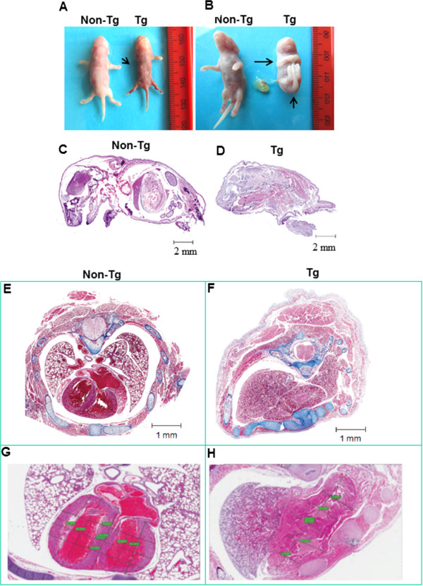
Morphological change in p49Tg mice versus non-Tg mice. A. Image of p49Tg newborn (Tg) versus Non-Tg newborn. The arrow indicates the p49Tg newborn missed a leg. B. The arrows highlight the contracture deformities of the limbs with additional webbing or non-delineated appendages in the p49Tg newborn. The tissue sections from C to H were stained with H&E staining. C and D. Longitudinal section of p49Tg newborn versus non-Tg newborn, which revealed that the spine in the M-p49Tg newborn was shifted around to the abdominal cavity from the back to the front (dorsal to ventral). E. Cross sections of non-Tg newborn. F. Cross section of Tg newborn with asymmetric thoracic cavity. Both the left and right lungs were unexpanded in the dead newborn the Tg mouse. G. Tissue section of non-Tg heart. H. Tissue section of p49Tg heart, which revealed malformed heart.
