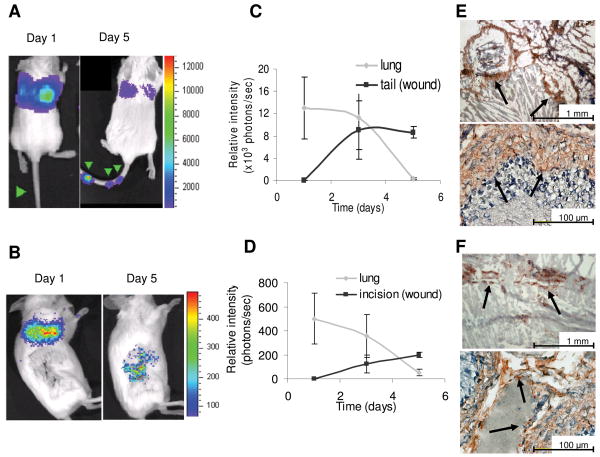
Figure 3.
hMSC co-localization with subcutaneous wound models. hMSC-ffLuc were injected immediately post needle puncture (Day 1) or 3 days post-surgical incision (Day 1). In the needle puncture model, hMSC-ffLuc were injected into SCID mice immediately post-wound infliction. Images are shown for representative animals at 1 and 5 days (A) post-needle puncture (n=5) and (B) post-lateral incision (n=3). Bioluminescent activity was quantified on days 1, 3, and 5, demonstrating a decrease in activity in the lung and concurrent increases of activity in the (C) tail wounds and (D) cutaneous incisions. IHC on sections of (E) tail and (F) wounded skin demonstrated incorporation of ff-Luc+ hMSC.

