Figure 5.
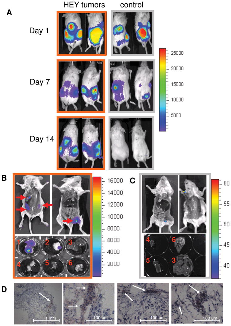
MSC tropism for HEY ovarian carcinoma. SCID mice were IP injected with HEY cells (n=3; orange outline) or PBS (n=3; grey outline). 15 days later, hMSC-ffLuc were IP injected in tumor-bearing and control mice (Day 1). (A) Images were acquired at days 1, 7, and 14 indicating initial dissemination throughout the peritoneal cavity, followed by specific localization in tumor-bearing animals and disappearance in control animals. On day 14, the mice were sacrificed bioluminescent activity was localized to sites of visible tumor development in the open cavities and dissected organs [(1) ventral tumor, (2) dorsal tumor, (3) liver, (4) kidney, (5) spleen, and (6) heart and lungs] of (B) HEY-bearing but not in (C) control mice. (D) IHC for ffLuc on tumor sections from the HEY-bearing mice confirmed the presence of hMSC (magnification as indicated).

