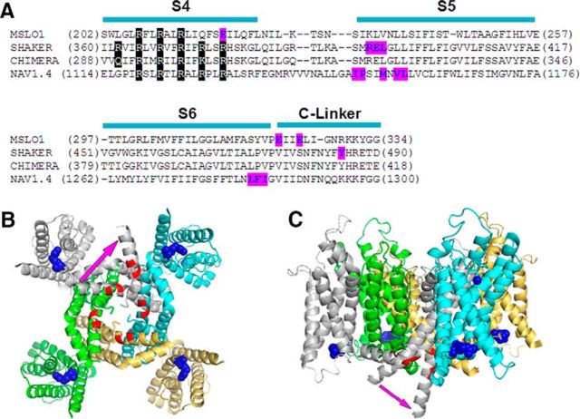
Figure 6.
A structure model of BK channels. A, Sequence alignment of BK, shaker (Batulan et al., 2010), Kv1.2/Kv2.1 chimera channels (Long et al., 2007), and sodium NAV1.4 channel domain III (Muroi et al., 2010). Purple represents residues that interact with neighboring subunits. B, C, Bottom (B) and lateral (C) views of the crystal structure of Kv1.2/Kv2.1 chimera channel (Long et al., 2007). Blue spheres represent residues corresponding to E219R in mSlo1; red represents the main chain of residues corresponding to E321/E324. Purple arrow indicates a possible S6 bending in mSlo1 channels.

