
Figure 10.
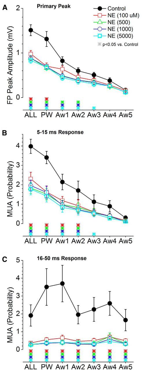
Population data measuring the effect of cortical noradrenergic activation on barrel cortex responses evoked by low-frequency whisker deflections. A, Primary negative peak amplitude of barrel cortex FP responses measured within a 5–30 ms window poststimulus during control and four doses of NE. The plot depicts the responses evoked by multiwhisker stimulation (6 whiskers simultaneously; ALL), the PW, and each of five adjacent whiskers (Aw1–Aw5) surrounding the PW. The color-coded asterisks at the bottom mark significant differences between control and each drug dose. B, Same as A, but MUA barrel cortex responses are measured during a short-latency (5–15 ms) window poststimulus. C, Same as A, but MUA barrel cortex responses are measured during a long-latency (16–50 ms) window poststimulus.