
Figure 4.
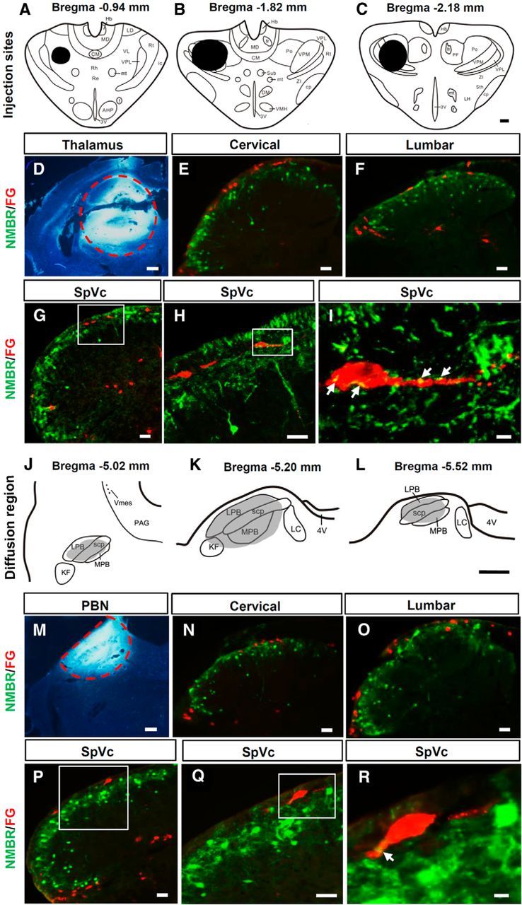
Retrograde tracing of NMBR+ neurons in the spinal cord and SpVc. A–C, Diagrams show FG injection sites (blacked areas) in the thalamus. D, The FG (bright white) injection site in the thalamus is indicated by a red dashed circle. E–H, There were no NMBR (GFP, green) and FG (red) double-labeled cells in the dorsal horn of the cervical spinal cord (E), lumbar spinal cord (F), and SpVc (G, H) in NMBR-eGFP mice. H, High-power image of the boxed area in G. I, High-power image of the boxed area in H showing that NMBR+ terminals (yellow) contact FG+ spinothalamic tract neurons. Arrows indicate contact sites. J–L, The shaded area indicates the diffused region of FG after PBN injection. M, Red dashed line defines the border of the injection site of FG in PBN. N–Q, Double staining in the dorsal horns of cervical spinal cord (N), lumbar spinal cord (O), and SpVc (P, Q) in NMBR-eGFP mice showed that NMBR+ (GFP, green) neurons were not FG+ (red) projection neurons to PBN. Q, High-power image of the boxed area in P. R, High-power image of the boxed area in Q showing that NMBR+ terminals made close contacts (yellow) with FG+ PBN projection neurons. Arrows indicate contact sites. Scale bars: A–D, J–M, 400 μm; E–H, N–Q, 40 μm; I, R, 10 μm. 3V, Third ventricle; 4V, fourth ventricle; AHP, anterior hypothalamic area, posterior; CM, central medial thalamic nucleus; cp, cerebral peduncle, basal part; DM, dorsalmedial nucleus; f, fornix; fr, fasciculus retroflexus; Hb, habenular nucleus; ic, internal capsule; KF, Kölliker-Fuse nucleus; LC, lateral cervical nucleus; LD, laterodorsal thalamic nucleus; LH, lateral hypothalamic nucleus; LPB, lateral PBN; MD, mediodorsal thalamic nucleus; MPB, medial PBN; mt, mammillothalamic tract; PF, parafascicular thalamic nucleus; Po, posterior thalamic nuclear group; Re, reuniens thalamic nucleus; Rh, rhomboid thalamic nucleus; Rt, reticular thalamic nucleus; scp, superior cerebellar peduncle; Sth, subthalamic nucleus; Sub, submedius thalamic nucleus; VL, ventrolateral thalamic nucleus; VMH, ventrolmedial hypothalamic nucleus; Vms, trigeminal mesencephalic nucleus; VPL, ventral posterolateral thalamic nucleus; VPM, ventral posteromedial thalamic nucleus; ZI, zone incerta.