
Figure 3.
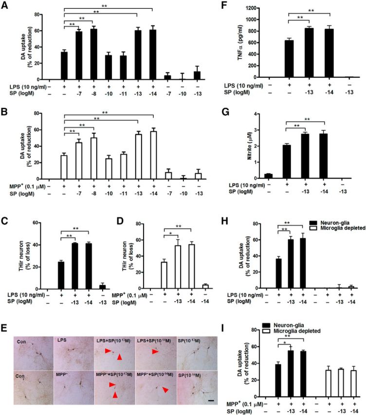
Exogenous SP potentiates dopaminergic neurodegeneration by activating microglia. A–G, Rat mesencephalic neuron-glia and microglia-depleted (H, I; microglia were removed from neuron-glia cultures by L-leucine methyl ester) cultures were treated with the indicated concentrations of SP and LPS (10 ng/ml) or MPP+ (0.1 μm). A, B, I, Seven days later, dopaminergic neurotoxicity was determined by [3H]-DA uptake assay and (C, D) concurrently quantified through immunocytochemistry for THir neuronal loss (n = 4–7) and (E) neurite atrophy (red arrowheads; n = 3). TNFα (F) and nitrite (G) levels were measured in the supernatant after 3 and 24 h of treatment, respectively. The results of [3H]-DA uptake and THir neuron counts are expressed as the percentage of reduced [3H]-DA uptake capacity and loss of THir cells (mean ± SEM, control group was considered as 0, no damage), respectively. The results of TNFα and nitrite are mean ± SEM. The data were analyzed using one- or two-way ANOVA followed by Tukey's post hoc test. *p < 0.05. **p < 0.01. Scale bar, 50 μm.