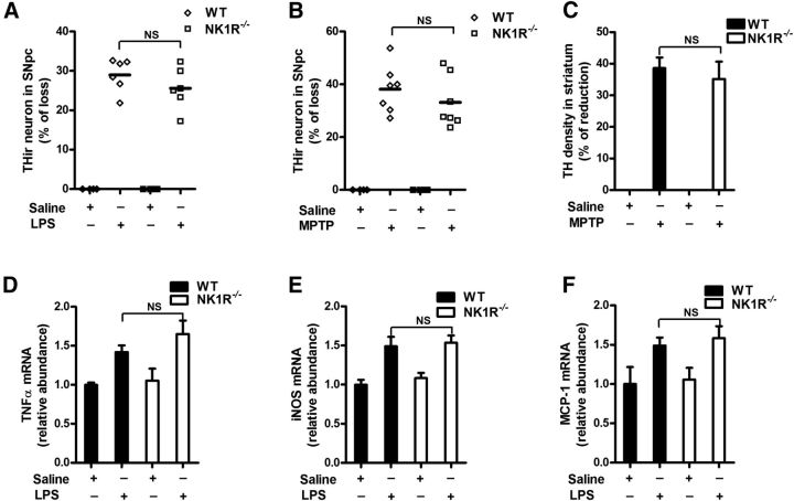
Figure 4.
NK1R deletion fails to reduce LPS/MPTP-induced nigral dopaminergic neurodegeneration and LPS-induced neuroinflammation in vivo. A single dose of LPS (15 × 106 EU/kg, i.p.) or a repeated MPTP regimen (15 mg/kg, s.c. for 6 consecutive days) was administered to NK1R−/− or WT mice. Ten months after LPS or 21 days after the last MPTP injection, the brains were collected. A, B, Nigral THir neurons in LPS-treated (A) and MPTP-treated (B) NK1R−/− and WT mice were counted stereologically. C, Striatal dopaminergic neuron fibers were quantified by TH density. D–F, Ten months after LPS injection, the mRNA levels of TNFα (D), iNOS (E), and MCP-1 (F) were determined in brains using RT-PCR. The results are expressed as the percentage of THir cell loss or density reduction (mean ± SEM, WT controls were considered as 0, no damage) in A–C and are expressed as a percentage of the controls (mean ± SEM) in D–F. The data were analyzed using two-way ANOVA followed by Tukey's post hoc test. n = 4–7. NS, Not significant.

