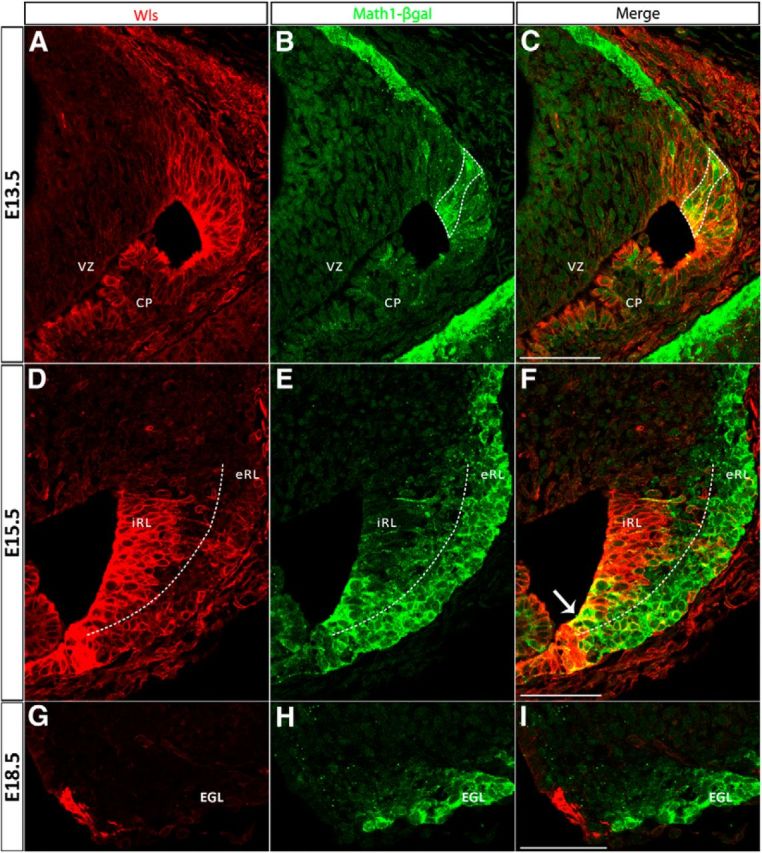
Figure 5.

The cerebellar RL progressively develops molecularly distinct populations during embryonic development identified by Wls and Math1 expression. The expression of Wls and Math1 was studied in Math1LacZ/+ mice, which express a β-gal reporter protein under control of the Math1 locus. Immunolabeling for Wls (red fluorescence, left) and β-gal (green fluorescence, middle) proteins is shown at E13.5 (A–C, top), E15.5 (D–F, middle), and E18.5 (G–I, bottom). At E13.5, Wls is expressed throughout the rhombic lip (A) and Math1 is strongly expressed in the emerging EGL and exhibits columns of expression in the RL (B). The overlap of Wls and Math1 expression domains in the cerebellar RL is shown in C (bounded by dotted line) where a subset of Math1-positive cells are also Wls positive at this early stage. Later at E15.5, Wls expression is localized predominately to the iRL (D), whereas Math1 expression is localized to the eRL and EGL (E). Expression at this time is largely nonoverlapping with only a few cells (arrow in F) coexpressing Wls and Math1 at the RL. At E18.5, the Wls-positive population found in the RL (G) is completely segregated from the Math1-positive population in the EGL (H). CP, choroid plexus; EGL, external germinal layer; VZ, ventricular zone. Scale bars: 50 μm.
