
Figure 2.
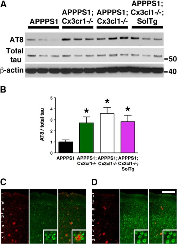
Hyperphosphorylation of MAPT in APPPS1 mice lacking membrane-anchored CX3CL1 signaling. A, Western blots of cortical lysates from APPPS1 (n = 6), APPPS1;Cx3cr1−/− (n = 5), APPPS1;Cx3cl1−/− (n = 4), and APPPS1;Cx3cl1−/−;SolTg mice (n = 4) at 4 months were probed with antibodies against phospho-MAPT (AT8), total MAPT, and β-actin. B, Quantification of band intensities revealed significant increases in AT8-reactive MAPT relative to total MAPT expression in APPPS1;Cx3cr1−/−, APPPS1;Cx3cl1−/−, and APPPS1;Cx3cl1−/−;SolTg genotypes compared with APPPS1 controls. C, D, Immunostaining brain sections for Aβ with monoclonal 4G8 antibody (red) and AT8 antibody (green) revealed pronounced phospho-MAPT accumulation within layer IV/V cortical pyramidal neurons of APPPS1;Cx3cl1−/− mice (D, insets), which was not observed in APPPS1 controls (C, insets). Scale bar, 100 μm; *p < 0.05, one-way ANOVA with Newman–Keuls post hoc test.