
Figure 4.
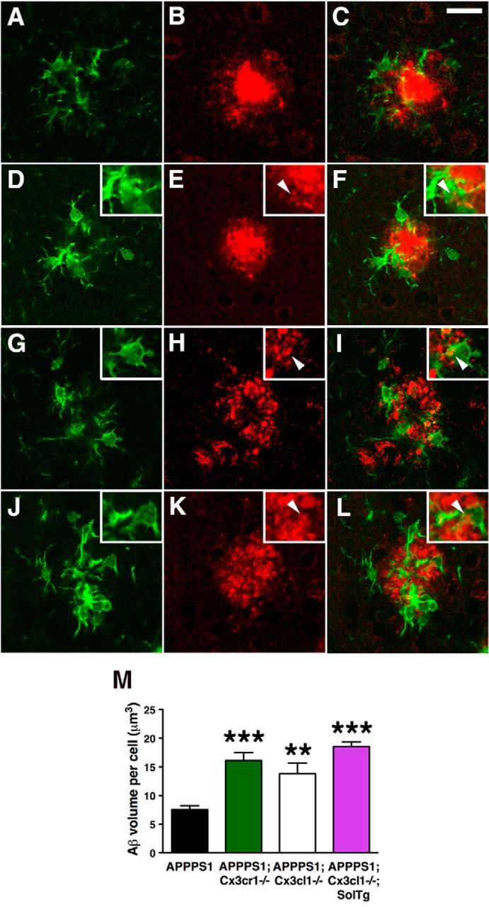
Enhanced microglial Aβ phagocytosis in APPPS1 mice lacking membrane-anchored CX3CL1 signaling. A–L, Brain sections from 4-month-old APPPS1 (A–C) and APPPS1;Cx3cr1−/− (D–F), APPPS1;Cx3cl1−/− (G–I), and APPPS1;Cx3cl1−/−;SolTg mice (J–L) were immunostained with the antibody against the microglial marker Iba1 (A, C, D, F, G, I, J, L, green) and the monoclonal Aβ antibody 4G8 (B, C, E, F, H, I, K, L, red). C, E, F, K, L, M, Quantification of Aβ phagocytosis (M) revealed increased intracellular Aβ volume per cell in APPPS1;Cx3cr1−/− (E, F, insets, arrowheads), APPPS1;Cx3cl1−/− (H, I, insets, arrowheads), and APPPS1;Cx3cl1−/−;SolTg mice (K, L, insets, arrowheads) compared with APPPS1 controls (C). Scale bar, 25 μm; **p < 0.01, ***p < 0.001, one-way ANOVA with Newman–Keuls post hoc test.