
Figure 5.
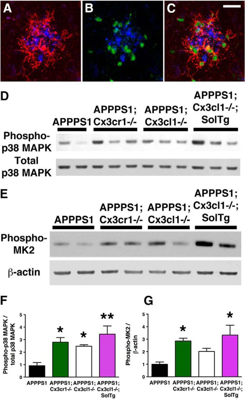
Increased p38 MAPK activation in APPPS1 mice lacking membrane-anchored CX3CL1 signaling. A–C, Immunostaining with the antibody against CD45, a marker for cells of hematopoietic lineage upregulated in activated microglia (A, C, red), the antibody against phospho-p38 MAPK (B, C, green), and the monoclonal Aβ antibody 4G8 (A–C, blue) revealed predominant localization of phospho-p38 MAPK within plaque-adjacent microglia in APPPS1 mice at 4 months (C). D, Western blots of cortical lysates from APPPS1 (n = 4), APPPS1;Cx3cr1−/− (n = 5), APPPS1;Cx3cl1−/− (n = 4), and APPPS1;Cx3cl1−/−;SolTg mice (n = 4) at 4 months were probed with anti-phospho-p38 MAPK and total p38 MAPK antibodies. F, Quantification of band intensities revealed increased phopho-p38 MAPK levels relative to total p38 MAPK levels in APPPS1;Cx3cr1−/−, APPPS1;Cx3cl1−/−, and APPPS1;Cx3cl1−/−;SolTg mice compared with APPPS1 controls. E, G, In addition, Western blot analysis of cortical lysates for phospho-MAPKAPK2 expression relative to β-actin (E) revealed significant increases in APPPS1;Cx3cr1−/− (n = 5) and APPPS1;Cx3cl1−/−;SolTg mice (n = 4) compared with APPPS1 controls (n = 6; G). Scale bar, 25 μm; *p < 0.05, **p < 0.01, one-way ANOVA with Newman–Keuls post hoc test.