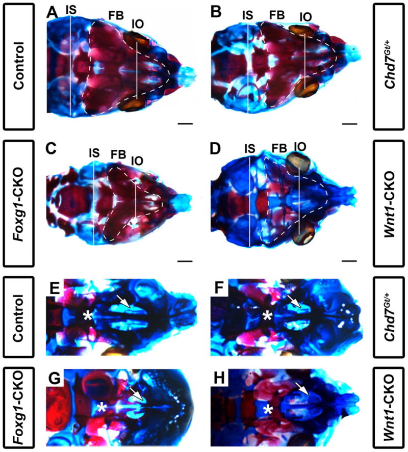
Figure 2.

Chd7 conditional mutants have shortened intraocular and intersquamosal distances, hypoplasia of the maxillary shelves, and dysplasia of nasal conchae. Skeletal preparations of postnatal day 1 control (Cre-negative, Chd7Gt/+ or Chd7f/f) (A,E), Chd7Gt/+ (B,F), Foxg1-CKO (C,G), and Wnt1-CKO (D,H) mice. Alizarin red and alcian blue staining show bone and cartilage, respectively. (A-D) are representative dorsal images of the calvaria while (E-H) are representative dorsal images of the developing maxillary shelves (with mandibles removed). Foxg1-CKO (C) mice exhibit shorter interocular (white line, IO; p<0.001, N=7) and intersquamosal (white line, IS; p<0.001, N=7) distances compared to control (A), Chd7Gt/+ (B), and Wnt1-CKO (D) mice. Foxg1-CKO (C) mice exhibit a visibly smaller frontal bone (FB) surface area (white, dotted line) relative to control (A), Chd7Gt/+ (B), and Wnt1-CKO (D) mice. Wnt1-CKO (D) mice lack frontal bone ossification (as demonstrated by alizarin red staining) relative to control (A), Chd7Gt/+ (B), and Foxg1-CKO (C) mice. In control (E) and Chd7Gt/+ (F) mice, appropriately formed palatal shelves (asterisk) and ventral nasal conchae (white arrow) are present. Foxg1-CKO (G) mice have hypoplastic palatal shelves which approximate the midline (asterisk) and absent ventral conchae (white arrow), whereas Wnt1-CKO (H) mice have severe palatal shelf hypoplasia (asterisk) and dysplastic ventral conchae (white arrow). Scale bars in A-D are 1 mm.
