
Figure 5.
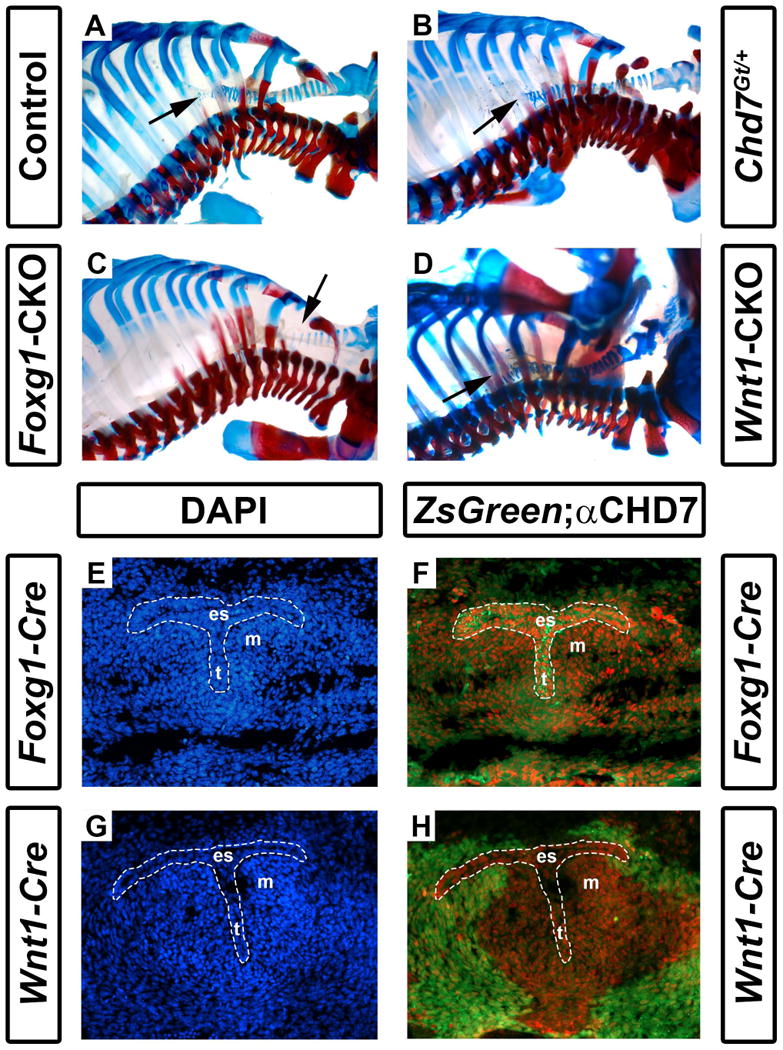
Foxg1-CKO mice have reduced numbers of cartilaginous tracheal rings. Shown are skeletal preparations of postnatal day 1 control (Cre-negative, Chd7+/f or Chd7f/f) (A), Chd7Gt/+ (B), Foxg1-CKO (C), and Wnt1-CKO (D) mice. Alizarin red and alcian blue staining reveal bone and cartilage, respectively. Normal numbers of tracheal rings (17-19) (arrows point to distal trachea) are present in control, Chd7Gt/+, and Wnt1-CKO mice, whereas Foxg1-CKO mice have fewer (11) tracheal rings. Panels E-H are transverse sections of Foxg1-Cre;ZsGreen (E, F) or Wnt1-Cre;ZsGreen (G, H) E11.5 embryos at the level of the laryngotracheal groove (indicated by the area within the dashed white lines) stained with DAPI (blue, labeling cellular nuclei) or anti-CHD7 (red). Green fluorescence (ZsGreen reporter) marks Cre-expressing cells and is overlaid with anti-CHD7 (red) in (F) and (H). Chd7 and Foxg1-Cre are highly co-expressed in the developing esophagus (es), trachea (t), and the surrounding mesenchyme (m), whereas Wnt1-Cre activity is restricted to the surrounding mesenchymal tissues and does not overlap with Chd7 expression in the presumptive esophagus or trachea.