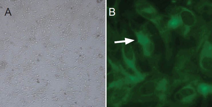
Figure 1.

Primary culture and identification of embryonic rat spinal nerve cells.
(A) Light microscopy (× 100) of primary spinal cord neurons, showing adherent and healthy neurons; some of the neurons showed small bumps. (B) Fluorescence microscopy (× 400) of spinal cord neurons identified using neuron-specific enolase, which fluoresced green (arrow) in the cytoplasm.
